Multibeam X-ray ptychography
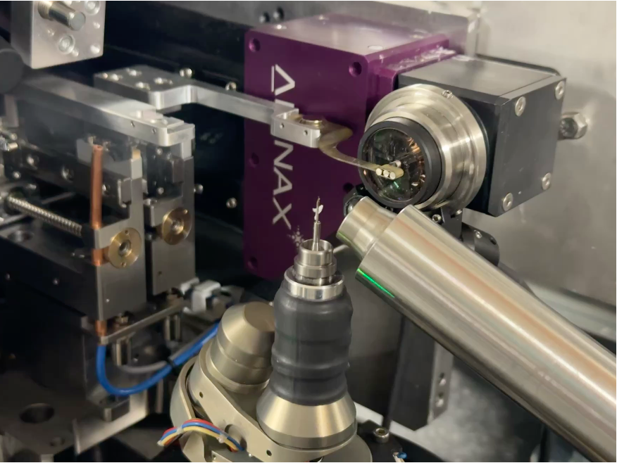
Ptychography needs coherent light, and at X-ray energies above 10 keV only a small part of a synchrotron beam is coherent; the rest goes unused. Multibeam ptychography splits the beam with an array of lenses into several beams that scan neighbouring regions of the sample at the same time, so the measurement speeds up in proportion to the number of beams.
Beam-scanning ptychography
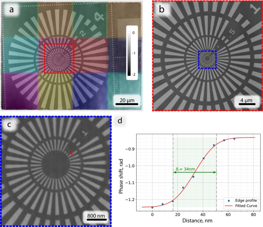
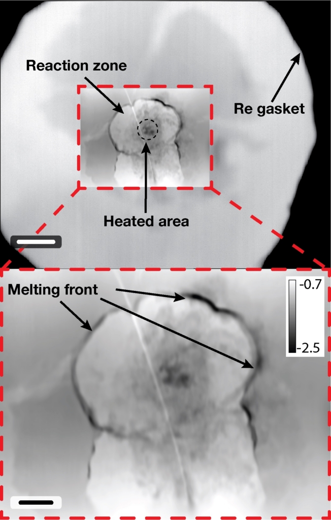
In conventional ptychography the sample moves through the beam with nanometre precision, which is hard when the sample sits inside a heavy or bulky cell. Instead we steer the beam across a sample that stays still, using a tilting mirror system, so the scanning speed no longer depends on the size or weight of the sample environment. In the first demonstration, at 12.3 keV, the method reached a resolution of about 40 nm and showed iron melting and oxidising inside a diamond anvil cell at a pressure of 50 GPa. The same scheme suits cryogenic, high-pressure and heated samples, and reactors that hold catalysts under working conditions.
Cryo X-ray ptychography
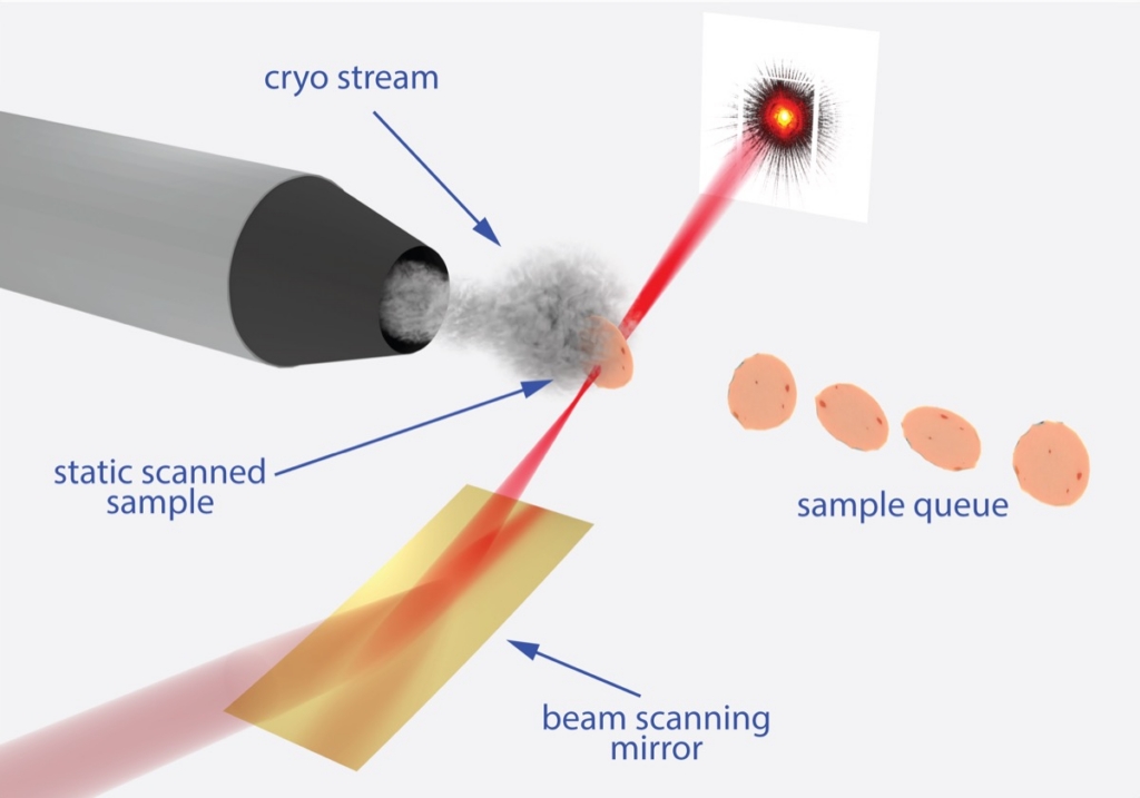
Biological tissue is usually stained with heavy metals and embedded in resin before it is imaged at the nanoscale, and both steps can change it. The team develops ptychography of frozen, hydrated specimens that are kept cold in a stream of cryogenic gas, so cells and tissues are seen in their natural state. Phase contrast makes unstained soft tissue visible with hard X-rays, and because ptychography needs no lens close to the sample, there is room for the cold gas stream around it.