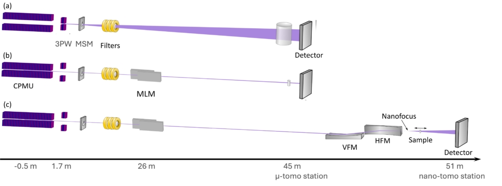
The beamline design includes two photon sources, a Cryogenically cooled Permanent Magnet Undulator (CPMU) and a 3-Pole Wiggler (3PW). The beam from the CPMU will enable fast microtomography of millimetre-sized samples with routine acquisition rates up to several tomograms per second. The beam from the 3PW will enable tomography of large samples up to ~ 45 mm x 4.5 mm (h x v), with no need for beam expansion optics. Both sources will allow ultrafast radiography with acquisition rate of up to 100 kHz.
A large set of filters and a multilayer monochromator, that can be moved in and out of the beam, will provide highly intense parallel beams for fast tomography at the µ-tomography end-station. For higher spatial resolution, TomoWISE will feature a nanotomography end–station with fixed-curvature, graded multilayer Kirkpatric-Baez mirrors, used in conjunction with the multilayer monochromator, focusing the beam down to below 250 x 250 nm2 size at 30 keV. This will act as a source for projection microscopy in a cone-beam geometry, in which spatial resolution and field of view can be continuously adjusted by moving the sample between the focal spot and the detector.
The simple but flexible optical design of TomoWISE, together with the unique double source solution, will allow the choice among the following configurations adapted to the needs of distinct materials science research questions:
- WB -Wide Beam for the study of large samples – 45 mm x 4mm
- HT- High Throughput tomographic microscopy – Robot arm
HS- High Speed for capturing dynamic processes – 20Hz 3D - HR- High Resolution for nano-characterisation- 250 nm @ 30, 45 keV
Tailored computational resources and user-friendly software for data acquisition, reconstruction, and analysis are crucial components of the beamline. Dedicated hardware and staff resources will be pivotal to build a steppingstone for robust, precise, and fast exploration of Big Data for academic and industrial users. This development will enable informed decisions during the measurement and likely lead to the shortening of the time between the experiment and publication or the impact on industrial processes. The computing infrastructure will be divided into two parts: one to handle the workflow from the detector to the reconstructed tomogram and one for data visualisation, segmentation, and quantitative analysis. Among these, the visualisation of the data during the experiment is of the highest priority, as repeatedly requested by researchers using tomographic methods.