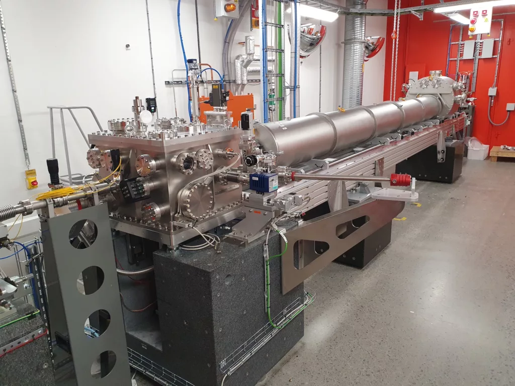
The imaging station (EH1) is the latest experimental endstation added to the NanoMAX beamline. It was opened to user operation in 2023. It provides an intense X-ray beam focused down to a 30 nm sized spot (the achievable smallest spot size is photon energy dependent). It is operated fully in vacuum with no windows between the sample and the detectors. A load-lock system allows to load up to 20 sample pins at once into the chamber. The endstation is designed for X-ray fluorescence mapping and ptychographic imaging in 2D and 3D.
Methods
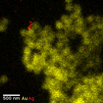
X-ray fluorescence mapping (XFM)
Using the nano-focused beam, information about the local elemental composition of the illuminated area can be extracted by measuring the secondary X-ray fluorescence photons using the energy-dispersive detectors mounted to both sides of the sample. This arrangement is especially advantageous for XRF tomography (3D) measurements.
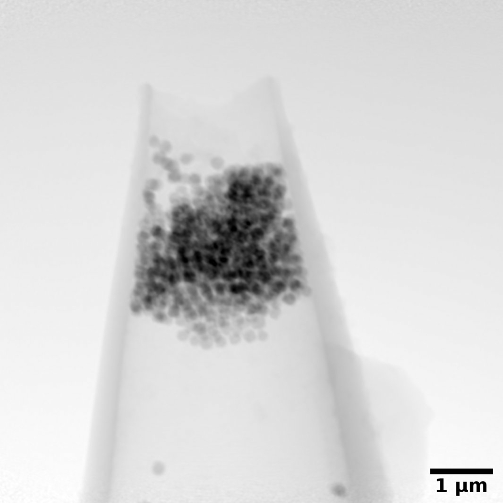
(Scanning) Coherent diffraction imaging (CDI) – ptychography
The beamline design allows to freely choose the degree of coherence in the probing beam, facilitating coherent diffraction imaging methods. These allow to measure the complex transmission function of the sample, as well as the complex beam profile. Both with a resolution better than the size of the focused beam. Again, 2D, as well as 3D measurements are possible.
Optics and beam size
The NanoMAX imaging endstation (EH1) uses a pair of Kirkpatrick–Baez (KB) mirrors to focus the X-ray beam into a tiny intense spot. These are the default optics offered to all users of the endstation. In the past Fresnel Zone Plates (FZPs) were used as focusing optics. FZPs are still an option for user operation, but their potential usage needs to be explicitly justified and agreed upon with the beamline scientists prior to proposal submission.
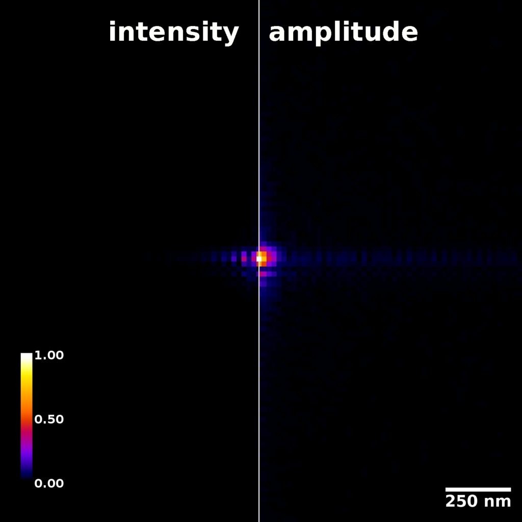
The KB mirrors at the imaging endstation provide a diffraction limited focal spot. At an incident photon energy of 12 keV they can focus a fully coherent X-ray beam down to a spot size of about 40 nm x 40 nm full width at half maximum (FWHM). A beam profile measured at the focus position shown in the figure to the right.
The mirrors are designed for an operation between 6 keV and 15 keV. While operation at higher photon energies is possible, it is not advisable, as the focal spot intensity will strongly diminish and the focal spot size will increase due to the KB mirrors (partially) reflecting at angles larger than the critical angle for total reflection.
Detectors
The NanoMAX imaging station offers two types of detectors.
- Firstly, a large in-vacuum pixel detector (EIGER2 X 4M) in the forward direction at roughly 7.1m distance from the sample for coherent diffraction imaging methods (ptychography).
- Secondly, two in-vacuum Silicon Drift Diode X-ray fluorescence detectors with carbon windows used for elemental mapping.
Planned developments
- Cryo-cooling to liquid nitrogen temperatures at the measurement position (in the X-ray beam) is planned, but has not been commissioned yet.
- Interferometric tracking of the sample position is planned to be added to the endstation.
- A publication on the details of the imaging endstation and its capabilities is currently in preparation.