Diffraction station parameters
| Photon energy | 5-28 keV | Routine operation 8-15 keV |
| Focal spot size | 50 – 200 nm Measured: 98 nm at 8 keV, 62 nm at 14 keV | Diffraction limited spot, the spot size is energy dependent. See doi.org/10.1364/OE.386068 |
| Focused beam divergence | Approximately 1.2 mrad (both vertical and horizontal) | energy independent |
| Coherent Flux | Fully coherent beam: 6×10¹⁰ at 8keV, 3×10¹⁰ at 10keV, 8×10⁹ at 14keV | The flux at the sample position depends on energy, see Beamline Optics and doi.org/10.1364/OE.386068 |
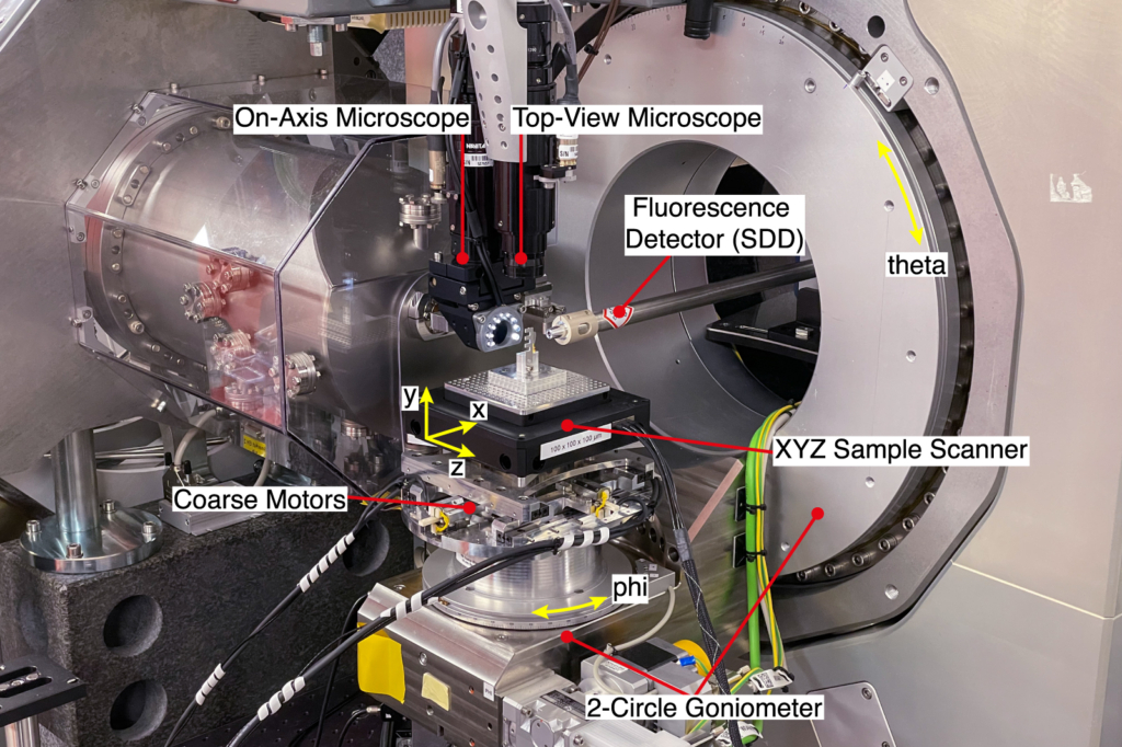
| 2-circle Goniometer | Theta: stroke [-5, 90] deg, resolution: 10-4 deg Phi: stroke [-180, 180] deg, resolution: 10-4 deg | Theta is horizontal and perpendicular to the X-ray beam. Phi is perpendicular to Theta and vertical for Theta=0. Note that the goniometer does not provide tomographic capabilities. |
| Coarse sample scanner (XYZ) | Stroke: 20 mm, Resolution: 100 nm | This is an in-house development |
| Fine sample scanner (XYZ) | Option 1 (standard): 100 μm stroke in all directions Option 2: 200 μm stroke in each horizontal direction, 100 μm vertical stroke | NPXY100Z100-135 This option provides the stiffest frame and the highest resolution NPXY200Z100-271 |
| Accessible space | Approx. 50 mm working space upstream of the X-ray focus. | Always contact the beamline staff to discuss non-standard sample holders or environments. |
| Continuous scanning | A single fine axis (X, Y, or Z) can be scanned continuously for fast mapping in 2D. | |
| Sample Microscopes | On Axis and Top Microscopes Maximum resolution: 5 μm Maximum Field of View: 0.3–4 mm | |
| X-Ray Fluorescence | RaySpec single-element SDD, coupled with an XSPRESS3 pulse processor | |
| In-line Detectors | Eiger2 X 1M in vacuum, 1028 x 1062 pixels2, 75 μm pixel size Pilatus3 X 1M, 981 x 1043 pixels2, 172 μm pixel size, 1000 µm thick Si sensor High-resolution Crytur camera, 2048 x 2048 pixels2, 600 nm effective pixel size | Detector distance given by flight tube, approx 1-3.8 m Detector distance minimum 160 mm Flexible placement downstream of the sample |
| Diffraction detector | Eiger2 X 500K, 1028 x 512 pixels2, 75 μm pixel size Optional: Merlin Si Quad, 512 x 512 pixels2, 55 μm pixel size | Mounted on the detector robot arm. Legacy detector, please contact beamline staff. |
| Detector robot | Positions the detector in a spherical coordinate system with polar and azimuthal angles approx. [-5, 90] degrees, and radius approx. [200, 1000] mm. | |
| Flight tubes | Forward vacuum tube housing the Eiger 3.5 m from the focus (modular in 1 m segments up to 4.5 m). Entry window 1.0 um Si3N4. Inflatable He tube for the Bragg geometry, cut to length. Entry and exit windows 6 um mylar. | |
| Other integrated equipment | Keysight B2985A electrometer ALBA electrometers PandABox acquisition card for digital and analog input SRS EC301 potentiostat Alemnis Nanoindenter | |
| Energy scanning | Manual energy changes are possible on a timescale of minutes. Image stacks can be collected as functions of energy, but XANES and EXAFS measurements are not possible. | |
| Off-line microscopy | Two optical microscopes are available for off-line sample inspection. One is a high resolution microscope (Olympus BX53M), the other a long distance stereo microscope (Olympus SZX16). A Hitachi SU1000 electron microscope is also available at the beamline. |