Macromolecular Crystallography
Synchrotron radiation plays a key role for structural biology, with macromolecular crystallography (MX) being one of the major technique, providing atomic-resolution structural information of macromolecules. At MAX IV, the BioMAX beamline supports a wide range of MX experiments.
MicroMAX opens up new possibilities in macromolecular crystallography by enabling time-resolved studies of structural dynamics on milli- and microsecond timescales. See below for an application example using High-Viscosity Extruder (HVE) for sample delivery and triggering by a laser with an image of structural conformation changes after illumination.
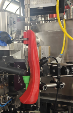
HVE with Laser Triggering
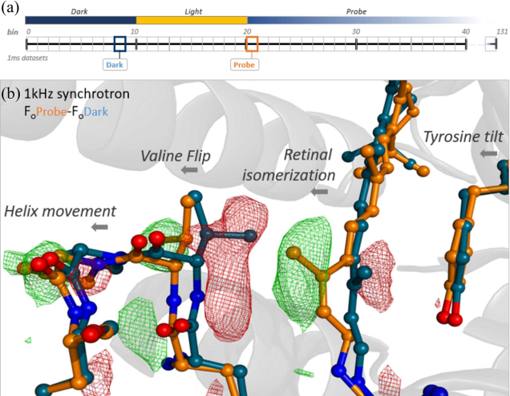
Example of Fo-Fo difference map: 1 ms after illumination minus dark. https://doi.org/10.1107/S2052252523008618
To support different triggering approaches and time ranges, several sample delivery methods have been and are being developed, including jet-based sample delivery (above), tape drive, fixed-target scanning and microfluidics.
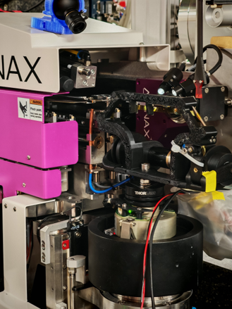
Tape drive (in development)
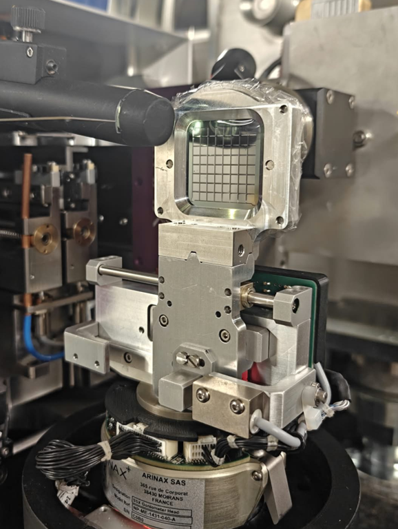
Fixed-target SSX scanning stage (in commissioning)
With a top-hat beam profile, rotation crystallography at MicroMAX supports radiation-damage studies and X-ray- or laser-triggered oscillation data collection, in addition to standard cryo-crystallography.
Cryo-Ptychography
Ptychography is a scanning coherent diffraction imaging (CDI) method capable of achieving nanometer-scale resolution. However, a major challenge in high-resolution X-ray imaging of biological specimens is radiation damage caused by the high X-ray dose required to achieve such resolution. This effect can be substantially mitigated by performing measurements under cryogenic conditions. At MicroMAX, we have adapted the cryo protein crystallography setup for cryo-ptychography, and recent feasibility studies have demonstrated resolution down to 50nm, with future prospects for three-dimensional tomography. See below for an example of an unfixed, unstained frozen cow embryo of ~150 µm diameter (5-6 days post-fertilization), measured with the sample cooled by a cold nitrogen gas stream, and the reconstructed image.
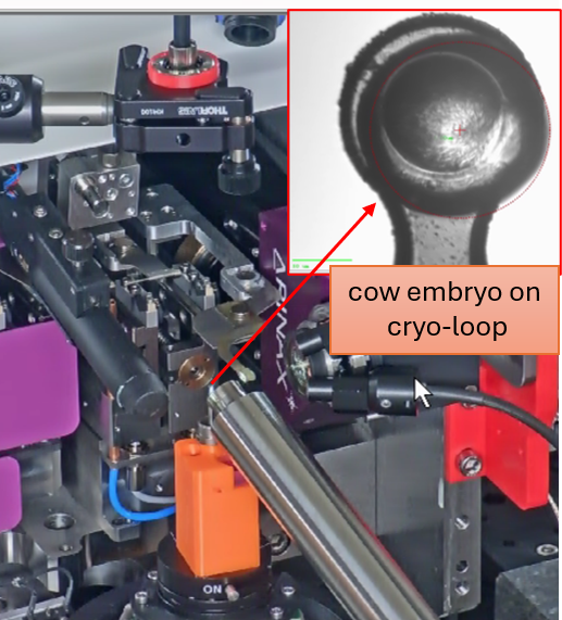
Experimental setup
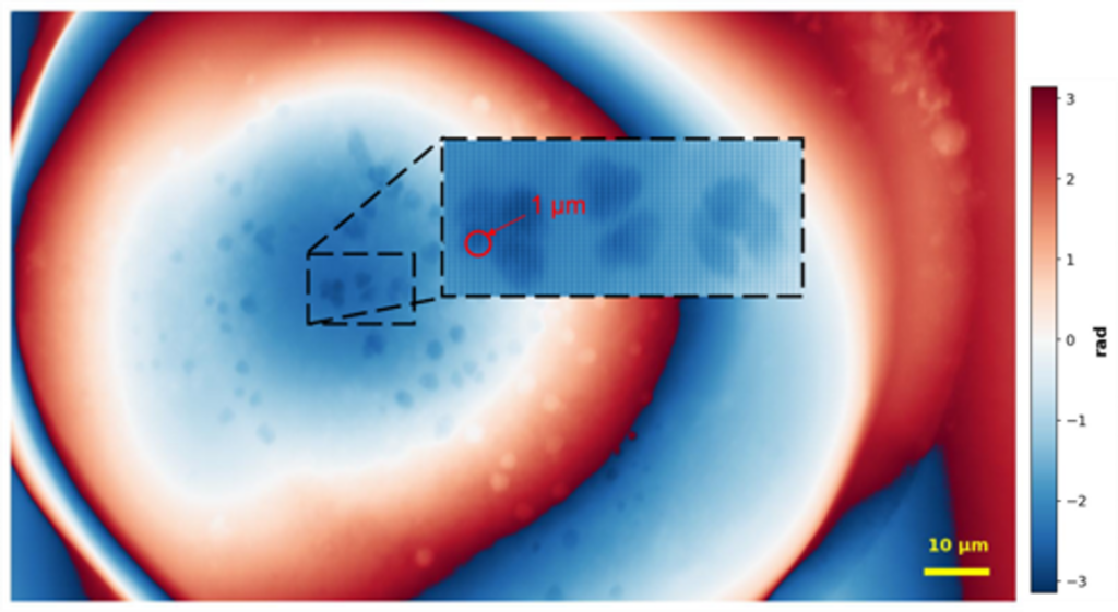
Reconstructed image (pixel size 34 nm)