Fast, high-resolution X-ray nanoscopy for samples too large for today’s methods
X-ray Nanoimaging Unit develops X-ray nanoscopy methods for large samples: many parallel beams that image large areas fast, a scanned beam for samples in heavy or bulky environments, and cryogenic imaging of unstained biological tissue. The methods build on the bright, coherent X-rays of the 3 GeV ring of MAX IV, the first fourth-generation synchrotron source.

Research lines
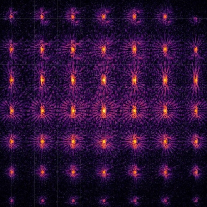
Multibeam X-ray ptychography. Several beams scan neighbouring parts of the sample at the same time, which speeds up the measurement without loss of resolution.
Beam-scanning ptychography. The beam moves across a sample that stays still, so samples in complex environments (cryogenic, high-pressure, heated) can be imaged at high resolution.
Cryo X-ray ptychography. Biological specimens are imaged frozen, in their natural state, without metal staining or resin embedding.
See more at Science & Techniques.
Projects and funding
HASTE: High-throughput multibeam ptychography supporting catalytic research. Röntgen-Ångström Cluster, 2022–2026. Develops multibeam ptychography for studies of catalysts at work.
MIRA: Beam-Steering X-ray Ptychography Supporting Cancer Research. Röntgen-Ångström Cluster, 2026–2030. Develops beam-steering ptychography to image cells for cancer research.
X-ray Hierarchical 3D Nanoscopy for large specimens. Swedish Foundation for Strategic Research (SSF), 2026–2029. Builds a nanoscope for hierarchical visualisation of the whole specimen down to the nanoscale.
Key publications
Keywords: X-ray ptychography · nanoimaging · X-ray microscopy · phase-contrast imaging · cryogenic imaging · catalysts · microchips · biological tissue · high pressure
- Yang, R., Villanueva-Perez, P., Li, T., Lyubomirskiy, M., Fevola, G., Falch, K. V., Sztucki, M., Kahnt, M. Optimizing probes for multi-beam ptychography. Optics Express 34(7), 12463 (2026). doi.org/10.1364/OE.582830
Compares ways to shape the beams and shows which make imaging with many beams most reliable. - Li, T., Falch, K. V., Garrevoet, J., Dubrovinsky, L., Lyubomirskiy, M. High-resolution ptychographic nanoimaging under high pressure with X-ray beam scanning. PNAS 122(43), e2514163122 (2025). doi.org/10.1073/pnas.2514163122
Scans the beam instead of the sample and images iron reacting at 50 GPa at a resolution below 50 nm. - Åstrand, M., Vogt, U., Yang, R., Villanueva Perez, P., Li, T., Lyubomirskiy, M., Kahnt, M. Multi-beam multi-slice X-ray ptychography. Scientific Reports 15, 9273 (2025). doi.org/10.1038/s41598-025-93757-0
First combination of multibeam and multi-slice imaging, for thick samples, shown at the NanoMAX beamline of MAX IV. - Li, T., Kahnt, M., Sheppard, T. L., Yang, R., Falch, K. V., Zvagelsky, R., Villanueva-Perez, P., Wegener, M., Lyubomirskiy, M. X-ray multibeam ptychography at up to 20 keV: nano-lithography enhances X-ray nano-imaging. Advanced Science 11(30), 2310075 (2024). doi.org/10.1002/advs.202310075
Twelve parallel beams from 3D-printed lenses image at 34 nm resolution, at 13 and 20 keV. - Lyubomirskiy, M., Wittwer, F., Kahnt, M., Koch, F., Kubec, A., Falch, K. V., Garrevoet, J., Seyrich, M., David, C., Schroer, C. G. Multi-beam X-ray ptychography using coded probes for rapid non-destructive high resolution imaging of extended samples. Scientific Reports 12, 6203 (2022). doi.org/10.1038/s41598-022-09466-5
Gives each beam its own phase code, so three beams image a microchip 2.7 times faster than one.