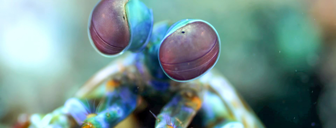
A group of Danish researchers have demonstrated three-dimensional sub-micrometre imaging of a whole, multi-millimetre sized, mantis shrimp eye. The eye was chosen to show the imaging opportunities of highly complex biological tissue at a fourth generation synchrotron.
The mantis shrimp is most well-known for punching prey using their club. They also have one of the most advanced eyes of all creatures in nature. Where humans have three channels for colour, red, green and blue, the mantis shrimp has sixteen. Apart from the usual colours of visual light, the shrimp can see both ultraviolet and polarised light as well. However, the mantis shrimp eye is still quite poorly understood by science. To study this type of tissue, fourth generation synchrotrons are very useful tools with enhanced contrast mechanisms and three-dimensional imaging capabilities.
For imaging the mantis shrimp eye and other biological tissue, a goal is to achieve a wide field of view and image the entire structure while maintaining high resolution in three dimensions. This way, it will be possible to “zoom” in on the image, and the microscopic structures can be understood within the context of the larger tissue.
“The most important result of our study was that we were able to image the entire eye in three dimensions. This allowed us to quantify specific structures within the eye, which paves the way for future simulation and modelling of the optical properties of these fascinating eyes,” say the researchers.
The imaging method opens the important possibility of gathering quantitative data and statistics that would not be possible in two dimensions. This allowed for studying the details of the muscles that move the eye and the photo filters, a pigment that is found before each unit of the compound like eye.
“We could see the size of the photo filters in the eye and quantify the spacing found in the periodic muscle structures, which is exciting as there is only sparse knowledge on muscle structure in invertebrates. We also discovered a vesicular network, which had not been found before in the mantis shrimp eye. This network weaves in between the cone cells [light receptor cells, editor’s note], and would thus be hard to capture with other methods. It was only possible to discover these features by imaging the eye in full with sub-micrometre resolution,” say the researchers.
The high level of coherence of the light in the fourth-generation synchrotron beam allows the use of not only the absorption but also the phase of the light as a contrast mechanism for three-dimensional imaging.
“The eye has very low contrast for the X-rays, and thus, imaging it places great demands on the quality of the light source used. Therefore, the high coherence of MAX IV – the first fourth generation synchrotron – was instrumental for the study. The lowest contrast is at the lenses and muscles, whereas the chitin cup [a hard material part of the eye, editor’s note] that holds the squishy part of the eye was relatively easy to image,” say the researchers.
To accomplish the goal of being able to image the whole structure while keeping the sub-micrometre resolution, the researchers had to use something called stitching. In tomography, the three-dimensional imaging method used, a wider field of view results in lower image resolution because the pixels of the detector have a fixed size. By imaging the sample part by part and stitching the incomplete pictures together, it is possible to circumvent the problem. The method used to do the stitching correctly can, however, be quite complex.
“The stitching method we used had been proposed in literature, but never by us or at MAX IV. The eye was measured with the explicit purpose of developing the stitching. If not for this experiment, DanMAX would not have had the stitching capabilities we have today,” say the researchers.
The method is an important addition to the imaging protocols offered at MAX IV.
“This gives future users the option of scanning samples multiple cubic millimetres in size at full sub-micrometre resolution, demonstrating the capability of scanning an organ at the cellular scale. Furthermore, the study showcases the high coherence of the source, demonstrating that we can gain new insights through fourth generation light sources. Finally, it demonstrated how imaging at DanMAX can provide high-resolution structural information on challenging biological targets and thus helps broaden the overall capabilities in biophysics and structural biology at the facility,” conclude the researchers.