Recent research in Nature Communications led by Aarhus University reports on the intriguing life history of narwhals, a species of Arctic marine mammal. The study reveals atomic-level details of the helical structure of the whale tusk. This knowledge of complex tooth construction advances understanding towards potential innovations in materials design.
Data from scanning powder X-ray diffraction (PXRD) measurements at MAX IV’s DanMAX beamline shows the hydroxyapatite nanoparticles growth patterns in the tusk, specifically in the texture and variations in the crystallite size and unit cell parameters.
“The DanMAX experiments help understand the details of how the mineralized parts change througout the sample,” said Mads Ry Jørgensen, study co-author and scientist at DanMAX beamline. “It was surprising to see how clearly the annual growth rings showed up in the crystallographic parameters.”
The new findings, part of the NordForsk project, enhance the body of knowledge on narwhals, including from prior analysis conducted at MAX IV by Aarhus University scientists on the chemical composition and biomineralization of the tusk, both important potential markers of accelerating climate change.
Researchers from Aarhus University in Denmark, Chalmers University of Technology in Sweden, and the Greenland Institute of Natural Resources contributed to the recent work. X-ray analysis was carried out at MAX IV Laboratory, the Swiss Light Source (SLS) in Switzerland, and the European Synchrotron Radiation Facility (ESRF) in France. Narwhal tusk samples were obtained with CITES (Convention on International Trade in Endangered Species of Wild Fauna and Flora) permits.
The power of PXRD is the ability to yield very detailed information about the atomic- and microstructure, however, normal PXRD lacks any real spatial resolution. By performing the PXRD experiment with a microscopic beam and systematically raster-scanning the sample it is possible to obtain detailed information in each point.
– Mads Ry Jørgensen
Excerpt paraphrase (Aarhus University press):
“The narwhal tusk assembles its macroscopic helix from building blocks with opposing twists”
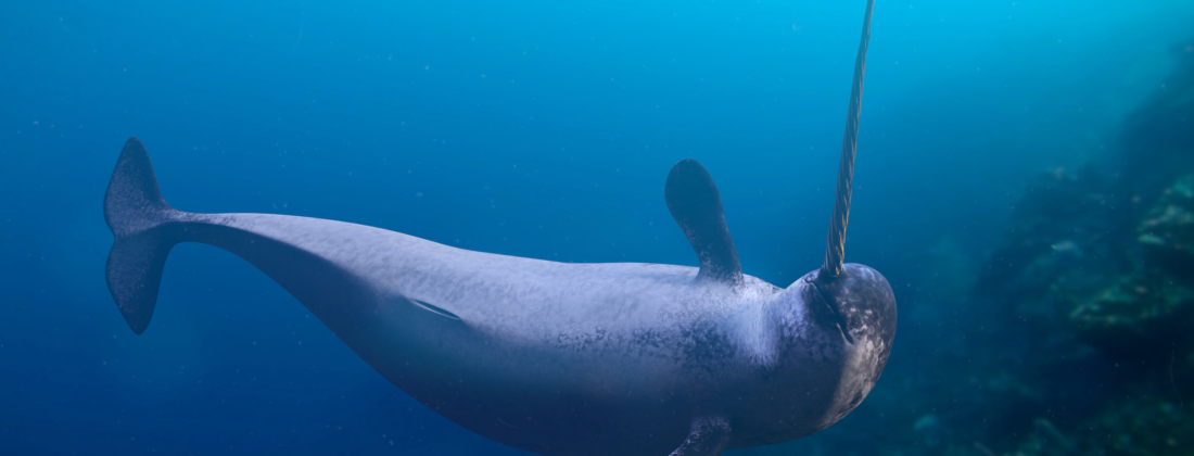
Narwhals – also known as the unicorns of the sea – are Arctic whales known for a single long tusk characteristic in males. The tusk reaches up to a third of the animal length and has an enigmatic left-hand helical structure that has puzzled researchers since the Middle Ages, when narwhal tusk material was sold as unicorn horn. The tusk was prized as a protection against poison, a prevalent medical myth of the era. How the helical structure is related to the structural arrangement of the building blocks of the tusk has remained an open question. This question has now been answered as reported in Nature Communications by Rodriguez-Palomo and colleagues.
The authors used X-ray 3D imaging techniques to uncover the hidden structure of the tusk. Using a technique called tensor tomography, they successfully mapped the orientation of tusk building blocks in three dimensions. Tensor tomography uses synchrotron radiation small-angle X-ray scattering (SAXS) signals from the mineralized collagen fibrils of the tusk to obtain structural information. Due to the high complexity and size of the tusk structure, the researchers combined the capabilities of three synchrotron facilities, MAX IV (Lund, Sweden), the Swiss Light Source (SLS) (Villigen, Switzerland), and ESRF (Grenoble, France).
The tusk is in fact the left tooth, consisting of dentine on the inside and cementum on the outside – unlike human teeth, there is no enamel. The authors found that the mineralized collagen fibrils follow a helical arrangement with opposite handedness in dentine and cementum, building two helices with opposite directions. This allowed them to determine the molecular origins of this fascinating case of macroscopic chirality. Remarkably, the overall helical arrangement was preserved across annual growth bands in the tusk that were imprinted in the tusk mineral nanocrystals.