Cryo-electron microscopy (cryo-EM) is a Nobel Prize–winning, cutting-edge technique for visualizing biological molecules in their near-native state at very high resolution. By rapidly freezing proteins and macromolecular complexes, Cryo-EM makes it possible to study their three-dimensional structures without the need for crystallization.
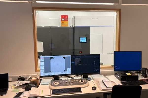
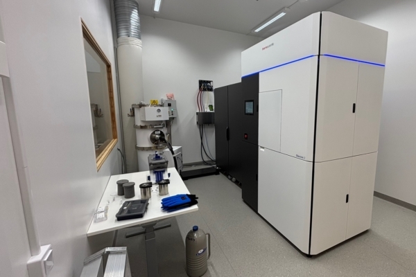
The Cryo-EM for Life Sciences Facility at Lund University provides access to a Thermo Fisher Glacios 2 transmission electron microscope (cryo-TEM), supporting both single particle analysis (SPA) and cryo-electron tomography (cryo-ET). Our facility also offers sample preparation services, including grid vitrification using a Thermo Scientific Vitrobot, and quality control by screening.The infrastructure is designed to support life science researchers at Lund University and beyond. We are also an infrastructure platform within SciLifeLab Lund.
For more information, please contact us at cryoem-facility@med.lu.se. We would love to discuss your project!
Key Principles
- Vitrification – Samples are rapidly frozen in liquid ethane, embedding biological macromolecules in a thin layer of vitreous ice. This preserves their native structure without chemical fixation or staining.
- Imaging – The frozen-hydrated sample is placed in a transmission electron microscope (TEM) and exposed to an electron beam. Direct electron detectors capture thousands of images at very low doses to minimize radiation damage.
- Image processing – Advanced computational methods align and combine two-dimensional projections to reconstruct a three-dimensional density map of the molecule.
- Model building – Atomic models can be fitted into the density maps, providing detailed structural insights.
Advantages
- Works with small sample amounts and heterogeneous complexes
- Captures multiple conformations and dynamic states
- Provides structural information close to atomic resolution
- Applicable to a broad range of targets, from soluble proteins to membrane proteins, viruses, ribosomes, and even cellular sections
Techniques
The facility supports imaging for:
- Single-particle analysis (SPA): High-resolution 3D structure determination of proteins and complexes.
- Cryo-electron tomography (cryo-ET): 3D imaging of macromolecules and cellular environments in their native state.
Applications
Cryo-EM @ LU enables research on:
- Membrane proteins and large macromolecular assemblies
- Viruses, ribosomes, and supramolecular complexes
- Mechanistic studies in molecular medicine and drug discovery
- Nanomaterials and soft matter
User Support
Our staff provide:
- Training in sample preparation, microscope operation, and data processing
- Assisted or independent use of Glacios 2
- Support in experimental design, optimization, and data analysis