Macromolecular Crystallography (MX) is an essential technique to determine the structure of biological molecules like proteins and nucleic acids to high resolution. 4th generation sources such as MAX IV, coupled with the use of advanced beamline instrumentation and fast readout detectors make it possible to study macromolecular crystals both by the conventional rotation method and explore the structural dynamics at a scale of milliseconds using the recently developed Synchrotron Serial Crystallography (SSX) methods.
The insights into Macromolecular Crystallography offers into the biological function of macromolecules have important applications in medicine, drug development and environmental science. MX methods are highly complementary with other structural biology X-ray techniques like BioSAXS and X-ray Spectroscopy, as well as Cryo-electron Microscopy and Neutron Diffraction, and they are a key tool in integrative structural biology.
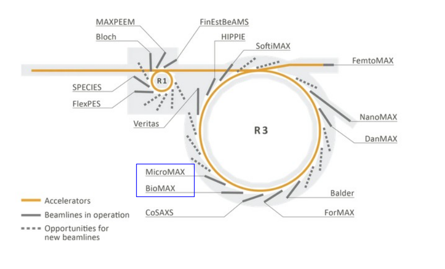
MX Facilities at MAX IV
At MAX IV there are two beamlines dedicated to Macromolecular Crystallography: BioMAX and MicroMAX.
- BioMAX is a versatile beamline, with high flux, variable beam focus and a large energy range. It is capable of a wide array of techniques, including high throughput automated oscillation experiments, de novo phasing and serial crystallography.
- MicroMAX is specialized in microfocus applications and time resolved experiments in the order of KHz. It has variable bandwidth, with flux of the order of 1013 – 1015 photons/s and a minimum focus of 5 x 5 μm2 which will be later reduced to 1 x 1 μm2.
The MX group at MAX IV also hosts a Fragment based drug discovery facility, FragMAX, providing simple workflows for large-scale crystal preparation, data collection and analysis.