Regular AFM
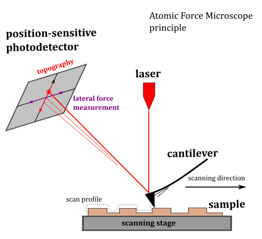
The Atomic Force Microscopy relies on scanning a very sharp tip across a surface of interest at a very close proximity or even touching it. The tip “sits” on a thin flexible stripe, usually metal-coated silicon, called a cantilever. The back of the cantilever is illuminated with the laser light, and the reflection is observed on a position-sensitive detector (PSD). Due to a large lever of the light path even smallest bends of the cantilever will result in a measurable reflection travel on the detector. Scanning the sample laterally against the cantilever and monitoring the respective displacement of the reflected light on the PSD, one obtains a height map or topography of the sample. The principle of Atomic Force Microscopy is illustrated below, left (clickable).
Peak Force and Quantitative Nanomechanical Mapping
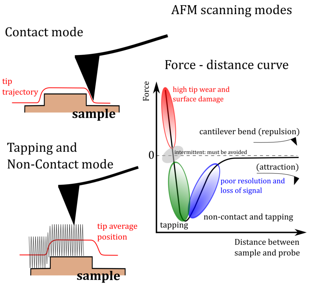
Peak Force(TM) scanning mode is a off-resonance tapping mode (the tip is driven at 2 .. 4 kHz) developed by Bruker, Inc., where at every cycle of tapping a force curve is being recorded and analyzed. This mode provides a precise and direct control of interaction between the tip and the surface, instead of an indirect resonance shift in regular tapping. Sampling force curves in every point of the scan allows for simultaneous extraction of such mechanical properties as sample’s stiffness, adhesion, dissipation and indentation. More information and examples to follow.
Infrared imaging and nanospectroscopy with AFM
more information to follow
Resonance-enhanced AFM-IR (contact)
coming soon
Tapping AFM-IR
coming soon