Researchers have found a biomaterial with surprising features in the skin of a lizard. The material is hard like enamel but is structured differently. Understanding the material on the nanoscale opens up new routes in designing for hard-wearing applications.
The Mexican beaded lizard has little hard plates in its skin called osteoderms, which are made of bone and topped with a so-called capping tissue. The plates protect the lizard from being hurt when bitten, but are also unique from a materials standpoint. An international research team has used the beamline DanMAX to study the material in the plates, particularly the capping tissue.
“We chose this particular lizard because previous work suggested it had a very stiff capping tissue. There are several open questions, such as how such a stiff tissue can form on top of bone and what the structure and mechanics of the capping material are,” says Henrik Birkedal, one of the contributors to the study.
The experiments show that the capping tissue is as hard as enamel. However, its internal structure is different. So, it looks like these types of hard materials could be realised in more than one way, and due to the variability in structure, potentially with different other mechanical properties besides the hardness.
“One of the most important results of the study was realising that nature fabricates hard mineralised tissues in a way that we had not seen before,” says Birkedal.
Researchers often study nature to understand and ultimately copy the materials created by evolution and natural selection. The research is called biomimicry or bioinspiration.
“Biomimicry and bioinspiration use nature as a source of inspiration to produce human-made methods, technologies, structures and new materials. Biology provides inspiration for what is at all possible – in the present case, the surprising finding of reaching enamel-like stiffness without the enamel-like ordered microstructure,” says Birkedal.
The research team imaged the structure and chemistry of slices from the osteoderms in two dimensions. They also imaged the osteoderms in 3D using X-ray tomography with phase contrast, which works much like a regular medical CT-scan but with much better contrast and resolution.
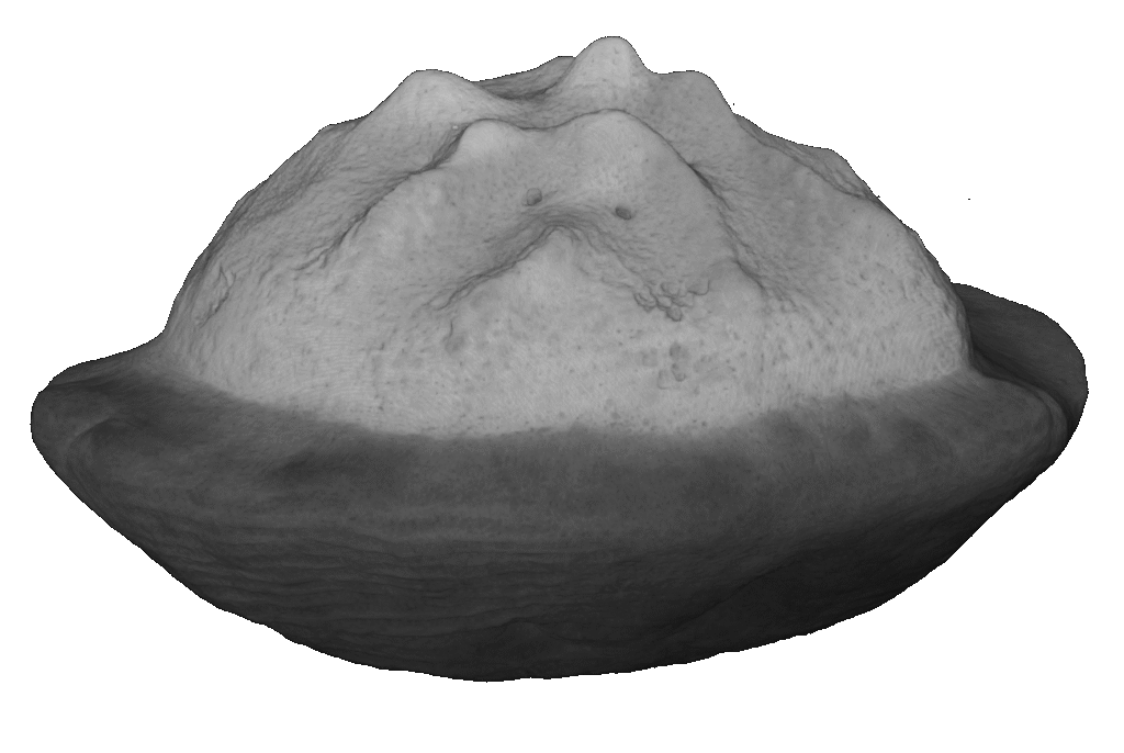
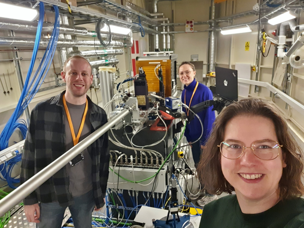
“DanMAX was the perfect place to conduct the experiment for multiple reasons. First of all, they have a good experimental station to perform X-ray diffraction and X-ray fluorescence simultaneously (methods for studying structure and chemistry, eds note), says Birkedal. “DanMAX has a very well-implemented experimental setup, which allows for a very user-friendly beamline control system that is very much appreciated. Finally, DanMAX allows for tomographic 3D imaging. This is the first publication of 3D imaging from DanMAX,” says Birkedal.
The researchers are planning to study more lizard skin in the future.
“The next step, which we are currently working on, is studying a larger range of lizards with the same bony structures in the skin. We want to know if their hard tissue shares the same building blocks and structure as other species or if it is an exceptional case among lizards. Regardless of the result, we will feel lucky that we came across a rare and unique case, and that we will help establish a new category of tissue that has been unknown to scientists until very recently,” says Birkedal.
Top image: A Mexican beaded lizard (Heloderma horridum), photo: Adobe Stock


