To capture extraordinary nanoscale details in crystallography takes the powerful coherent flux of a 4th generation light source. Recent work in Light: Science & Applications by an international research team has revealed 3D images of a complex crystalline star structure using Bragg ptychography and new advanced analysis tools at MAX IV’s NanoMAX beamline. The results demonstrate the possibility of unprecedented data quality beyond experimental limitations from new synchrotron sources.
It is the high brilliance of 4th generation synchrotrons which now makes high resolution 3D Bragg ptychography especially valuable for investigation of crystal samples, from biominerals found in teeth, bones, shells and more, to a diversity of technologically relevant materials exhibiting magnetic, ferro-electric, topological properties to cite a few.
“New microscopy tools can provide not only sharper images but allow completely new ways of studying extremely complex materials, improving our understanding of the world around,” said Dina Carbone, MAX IV Scientist and study author. “This is the first step to produce technologies that truly responds to our needs in an efficient and sustainable way.”
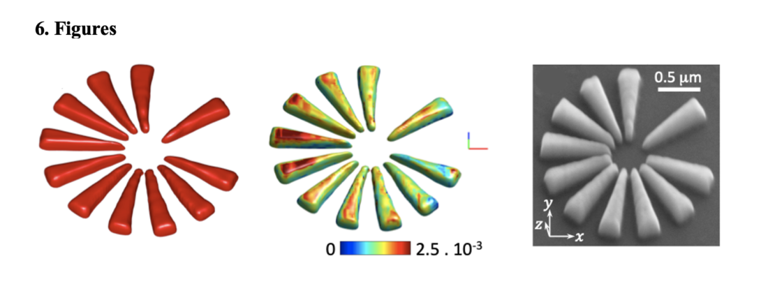
The current study succeeded in producing a 3D image of the silicon crystalline sample with internal atomic deformations. The star is a well-known structure, chosen to assess the capabilities of the new diffraction end-station of NanoMAX previously designed by Carbone. The research team involved pioneered the 3D Bragg ptychography technique in 2011, and continues with its development.
The experiment’s sharp, quantitative imagery is primarily attributed to the unprecedented flux of coherent X-rays impinging on sample. The large datasets produced surpass experimental limitations such as positioning errors or poor scanning conditions to obtain a high-resolution image of the sample. These circumstances are not possible to recreate at 3rd generation facilities, according to the study.
“Intensity was a crucial component of this study, but not the only one. Appropriate data processing and image reconstruction routines had to be developed to reach this image quality, which took quite some effort,” said study author Peng Li, now at Diamond Light Source in the United Kingdom.
These advances make Bragg ptychography compatible with a wider range of experimental set-ups at new synchrotron sources.
“Now it is time to strengthen the experimental and analytical competences of the user community interested in using such powerful microscopies. And this is a challenge and a responsibility of the light source facilities,” said Carbone.
“We will be happy to help with implementing this microscopy and make it available for a larger scientific community,” said Chamard.
The researchers are continuing their investigations with 3D microscopy at 4th generation sources, in the field of functional magnetic crystals and to investigate the intriguing structural properties of biominerals.