The powerful imaging techniques available at synchrotrons like MAX IV can work in tandem with medical imaging, enabling clinicians to offer cutting-edge care to their patients.
That was the takeaway from the Health session at MAX IV’s 37th user meeting. Organised by Arash Panahifar and Selma Maric, the session featured speakers from Lund University (LU), the European Molecular Biology Laboratory (EMBL), Rigshospitalet, and the Technical University of Munich (TUM).
““We want to harness the full power of MAX IV to enable rapid, 3D, multi-scale tissue characterisation,” Maric said. “Developing this methodology is about more than technology. It’s about giving clinicians better tools, and patients earlier, more accurate diagnoses. At MAX IV, our beamline scientists engage with an extraordinary diversity of samples and scientific challenges, and that breadth allows us to translate expertise across fields and drive novel solutions in emerging medical applications.”
Elizabeth Duke (EMBL) described applications of the high throughput tomography workflow her group has developed at the P14 structural biology beamline at PETRA III, which allows for highly automated data collection. This fast, non-destructive imaging can be deployed in projects such as the prediction and early detection of kidney transplant rejection. The correlation of the X-ray tomography images with lower resolution clinical imaging and higher resolution electron microscopy data has the potential to help clinicians identify what signs to look for to catch rejection early.
Looking ahead to the future of synchrotron imaging, Duke said that “multimodal, multi-length scale correlative imaging is the way forward to answer biological questions.”
Hanna Isaksson (LU) shared her research into how knee tissues move and change under pressure using time-resolved phase contrast X-ray tomography. This method allows researchers to see the changes as they occur, and in this case, allowed Isaksson to study the changes as mechanical strain was added to the connective knee tissue samples.
Julia Herzen’s (TUM) work has a more quantitative approach, using synchrotron imaging to study how many cells are affected by a given change by comparing morphology. Increased coherence at synchrotrons will help make this work more precise by allowing sharper imaging of small changes, and thus marking differences more clearly.
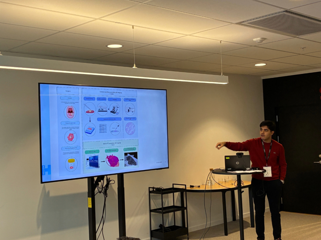
Ahmed Al Khafaf, a surgeon at Rigshospitalet in Copenhagen, said that there is a lack of 3D imaging of surgical specimens, like tumours. Pathohistology of biopsies can take days of laboratory work, while advanced X-ray imaging can produce results in a few hours. In the future, the time could be cut down to minutes. Implementing fast and precise 3D imaging could offer huge benefits in optimising tumour assessment.
The first step in Al Khafaf’s journey toward synchrotron imaging was microtomography at the Technical University of Denmark, which provides images within in hour. Phase contrast X-ray tomography at DanMAX beamline has further developed understanding of tumour margins.
“Once the entire workflow is optimized, including specimen preparation, image acquisition, and post-scan 3D rendering, the full potential of MAX IV will be unlocked,” Al Khafaf said. “The future will be to provide 3D imaging of surgically removed specimens – an approach that is not currently implemented. I believe MAX IV will be a key factor in this development.”
Beyond MAX IV, other consortia are already forming around the goal of exploiting X-ray synchrotrons as a real medical tool with immediate benefits for patients. Integrated X-ray and biomedical imaging (IXBI), an application for a Swedish Research Council excellence cluster, led by MAX IV and SciLifeLab, aims to develop an end-to-end pipeline for studying biopsies. This work unites clinical imaging with X-ray imaging and tomography at MAX IV and volume electron microscopy and spatial omics at SciLifeLab, analysed by AI-powered tools from SciLifeLab.
Ultimately, it will be the patients who benefit most from the rapid tissue characterisation and early diagnoses that X-ray synchrotrons can help provide.